OCT For Macular Degeneration: Wet Vs Dry Explained
Key Takeaways
- OCT (Optical Coherence Tomography) is a non-invasive imaging test that detects macular degeneration early.
- There are two types of macular degeneration: wet and dry.
- Wet macular degeneration is caused by abnormal blood vessel growth, while dry AMD is characterized by drusen deposits.
- The REVO-OCT Wellness Scan employs AI technology to enhance the accuracy of OCT scans.
- Regular OCT scans, such as those offered by Eyecare Opticians, help with monitoring the progression of AMD and adjust treatments accordingly.
Eyecare Opticians: Your Local Vision Care Experts
Discover quality vision care at Eyecare Opticians in Ham, Kingston Upon Thames.
Led by Consultant Optometrist Nish, with over two decades of diverse optometric experience, we offer cutting-edge eye care solutions tailored to your needs.
Our services include advanced diagnostic tests like Retinal Photography Optomap and Optical Coherence Tomography, alongside specialized treatments for Myopia control and Dry Eyes. Rated 4.9 stars on Google, we're committed to exceeding patient expectations with the latest equipment and personalized care.
Schedule your comprehensive eye exam today!
Introduction to OCT for Macular Degeneration
Macular degeneration is a serious eye condition that can lead to vision loss if not detected early. Fortunately, advances in technology like Optical Coherence Tomography (OCT) have made it easier to diagnose and manage this condition.
OCT is a non-invasive imaging test that utilizes light waves to take cross-sectional pictures of the retina, enabling eye doctors to see each of its distinctive layers and map and measure their thickness. These measurements help with diagnosis and provide treatment guidance for glaucoma and diseases of the retina, such as macular degeneration and diabetic eye disease.
The Importance of OCT in Eye Health
OCT allows optometrists to detect eye conditions like macular degeneration early, often before symptoms appear - early detection means early treatment, which can slow or even stop the progression of the disease. This is particularly important for AMD, which can lead to severe vision loss if left untreated.
- non-invasive and quick
- provides detailed images of the retina
- helps in early detection and monitoring of eye diseases
What is Macular Degeneration?
Macular degeneration, often referred to as age-related macular degeneration (AMD), is a common eye condition among people aged 50 and older. It causes damage to the macula - a small spot near the center of the retina that is responsible for sharp, central vision, needed for activities like reading and driving.
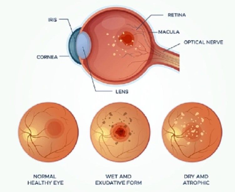
Wet Macular Degeneration
Definition and Key Characteristics
Wet macular degeneration, also known as neovascular AMD, is the less common but more severe form of age-related macular degeneration. It occurs when abnormal blood vessels grow under the retina and macula. These blood vessels can leak fluid or blood, leading to rapid and severe vision loss.
Formation of Abnormal Blood Vessels
In wet AMD, the formation of abnormal blood vessels is called choroidal neovascularization (CNV) - these new blood vessels are fragile and can cause the macula to lift from its normal position. This distorts vision and can lead to the formation of scar tissue, further deteriorating sight.
Symptoms of Wet AMD
Wet AMD symptoms can appear suddenly and worsen quickly. Key signs to look out for include:
- Visual distortions, such as straight lines appearing wavy
- Reduced central vision in one or both eyes
- A well-defined blurry spot or blind spot in your field of vision
- Colors appearing less bright
Risks and Progression of Wet AMD
Wet AMD progresses rapidly and can cause significant vision loss within a short period. Risk factors include age, genetics, smoking, high blood pressure, and obesity. If left untreated, wet AMD can lead to permanent central vision loss.
Treatment Options for Wet AMD
While there is no cure for wet AMD, several treatments can help slow its progression and preserve vision:
- Anti-VEGF Injections: These medications inhibit the growth of abnormal blood vessels.
- Photodynamic Therapy: A two-step treatment that uses a light-activated drug to destroy abnormal blood vessels.
- Laser Surgery: A high-energy laser is used to seal off leaking blood vessels.
Dry Macular Degeneration
Definition and Key Characteristics
Dry macular degeneration is the more common form of AMD, accounting for about 80-90% of cases. It progresses more slowly than wet AMD and occurs when the macula thins over time as part of the aging process, leading to gradual vision loss.
Presence of Drusen Deposits
An early sign of dry AMD is the presence of drusen - yellow deposits under the retina. While small amounts of drusen may not cause vision problems, larger and more numerous drusen increase the risk of developing advanced AMD.
- Small drusen: Generally do not affect vision.
- Intermediate drusen: May cause some vision changes.
- Large drusen: Significantly increase the risk of vision loss.
Symptoms of Dry AMD
Symptoms of dry AMD develop gradually and can include:
- Blurriness in the center of your vision
- Difficulty recognizing faces
- Need for brighter light when reading or doing close-up work
- Colors appearing less vivid
Risks and Progression of Dry AMD
- Age: Most common in people over 60.
- Genetics: Family history increases risk.
- Lifestyle: Smoking and poor diet can contribute to AMD.
Dry AMD progresses in three stages: early, intermediate, and late. Early detection through regular eye exams is crucial to manage the disease effectively.
Treatment Options for Dry AMD
While there is no cure for dry AMD, certain measures can help slow its progression and preserve vision:
- AREDS Supplements: High-dose vitamins and minerals can reduce the risk of advanced AMD.
- Healthy Diet: Consuming leafy greens, fish, and nuts can support eye health.
- Regular Eye Exams: Monitoring changes in your vision helps catch progression early.
Role of OCT in Diagnosing Macular Degeneration
OCT plays a crucial role in diagnosing and managing both wet and dry macular degeneration - the technology provides detailed images of the retina, allowing eye doctors to detect early signs of AMD and monitor its progression.
Benefits of Using OCT in Eye Examinations
- Early detection of retinal diseases
- Accurate measurement of retinal thickness
- Non-invasive and painless procedure
- Quick and efficient imaging
OCT Imaging in Wet AMD
In wet AMD, OCT can detect fluid or blood under the retina, indicating the presence of abnormal blood vessels. The device also monitors the effectiveness of treatments like anti-VEGF injections, allowing doctors to adjust the treatment plan as needed.
Managing Macular Degeneration with OCT
Managing macular degeneration effectively requires regular monitoring and timely adjustments to treatment plans, and OCT plays a pivotal role in this ongoing process. By rendering detailed images of the retina, OCT helps eye doctors track the progression of the disease and make informed decisions about treatment.
For instance, in wet AMD, the presence of fluid or blood detected by OCT may indicate the need for more frequent anti-VEGF injections.

Regular Eye Check-Ups: What to Expect
Here's what you can expect during a regular eye check-up:
- Review of your medical history and any changes in your vision
- Comprehensive eye examination, including visual acuity and retinal imaging
- Discussion of OCT scan results and any necessary adjustments to your treatment plan
- Recommendations for lifestyle changes or supplements to support eye health
Choose Eyecare Opticians for Age-Related Macular Degeneration Control
At Eyecare Opticians, we are committed to providing the highest level of care for patients with age-related macular degeneration. Our state-of-the-art REVO-OCT Wellness Scan uses AI-powered technologies like denoising and motion correction to enhance the quality and reliability of OCT scans. As such, we can detect macular degeneration early and monitor its progression with unparalleled accuracy.
Our experienced team of eye care professionals is dedicated to helping you preserve your vision and maintain your quality of life. We offer personalized treatment plans tailored to your specific needs and regular follow-up appointments to ensure that your condition is managed effectively.
Contact us to learn more about how to preserve your vision and manage macular degeneration effectively with advanced diagnostic tools and personalized treatment plans at Eyecare Opticians.
Frequently Asked Questions (FAQ)
What is the primary difference between wet and dry macular degeneration?
Wet AMD involves the growth of abnormal blood vessels under the retina, leading to rapid vision loss. Dry AMD, on the other hand, is characterized by the gradual thinning of the macula and the presence of drusen deposits, resulting in slower vision loss.
Can OCT detect early signs of macular degeneration?
Yes, OCT can detect early signs of macular degeneration - by providing detailed images of the retina, OCT can reveal changes in the retinal layers and the presence of drusen deposits, which are early indicators of AMD. Early detection through OCT allows for timely intervention and better management of the condition.
Is there a cure for macular degeneration?
Currently, there is no cure for macular degeneration; however, several treatments can help slow its progression and preserve vision. These include anti-VEGF injections for wet AMD and AREDS supplements for dry AMD. Regular eye check-ups and a healthy lifestyle can also help manage the condition.
How often should one get an OCT scan?
The frequency of OCT scans depends on the individual's risk factors and the stage of macular degeneration.
For those at risk or diagnosed with AMD, it is generally recommended to have an OCT scan at least once a year.
However, your eye doctor may recommend more frequent scans based on your specific condition and treatment plan - contact us at Eyecare Opticians and we’ll be happy to help with a personalised eyecare plan!
Can lifestyle changes help manage macular degeneration symptoms?
Yes, lifestyle changes can help manage macular degeneration symptoms and slow its progression. Some recommendations include:
- Eating a diet rich in leafy greens, fish, and nuts
- Taking AREDS supplements as recommended by your doctor
- Quitting smoking
- Maintaining a healthy weight and blood pressure
- Protecting your eyes from UV light by wearing sunglasses
By making these changes, you can support your eye health and reduce the risk of worsening macular degeneration.