Optomap For Retinal Detachment Diagnosis & Treatment: How It Works
Key Takeaways
- Retinal detachment symptoms include sudden flashes of light, floaters, and a shadow over the field of vision.
- Early detection of retinal detachment can prevent severe vision loss.
- Optomap retinal imaging captures over 80% of the retina in a single panoramic image.
- The method is non-invasive and doesn't require pupil dilation, making it more comfortable for patients.
- Optomap retinal scans, like those offered by Eyecare Opticians, are quicker and more accurate compared to traditional methods like standard eye exams and ultrasounds.
Eyecare Opticians: Your Local Vision Care Experts
Discover quality vision care at Eyecare Opticians in Ham, Kingston Upon Thames.
Led by Consultant Optometrist Nish, with over two decades of diverse optometric experience, we offer cutting-edge eye care solutions tailored to your needs.
Our services include advanced diagnostic tests like Retinal Photography Optomap and Optical Coherence Tomography, alongside specialized treatments for Myopia control and Dry Eyes. Rated 4.9 stars on Google, we're committed to exceeding patient expectations with the latest equipment and personalized care.
Schedule your comprehensive eye exam today!
What is Retinal Detachment?
Retinal detachment happens when the retina pulls away from its normal position. This is a medical emergency that needs immediate attention; the longer the retina remains detached, the less likely it is that vision can be restored.
if not treated quickly, it can lead to permanent vision loss or even blindness. Unfortunately, many people ignore the early symptoms, leading to more complicated and less effective treatments later on.
Symptoms to Look Out For
- Sudden flashes of light
- New floaters in your vision
- A shadow or curtain over part of your field of vision
Causes and Risk Factors
- Severe nearsightedness
- Previous eye surgery or trauma
- Family history of retinal detachment
- Aging, as the retina can thin and tear over time
Traditional Methods of Diagnosing Retinal Detachment
Standard Eye Exam
During a standard eye exam, an eye doctor uses a special magnifying lens to examine the retina. This method can identify tears and detachments, but it requires a highly skilled practitioner and doesn't always provide a complete view of the retina.
Pupil Dilation
Pupil dilation involves using eye drops to widen the pupil, allowing the doctor to see more of the retina - however, while effective, this method can be uncomfortable and time-consuming (the dilation process takes usually about 15 to 30 minutes for the drops to take full effect, which can make the entire eye exam more cumbersome for the patient). The procedure also makes the eyes sensitive to light for several hours afterward.
Ultrasound and Other Imaging Techniques
Ultrasound imaging is another method used to diagnose retinal detachment: it involves using sound waves to create an image of the inside of the eye. This method can be particularly useful when there is a lot of blood or other opacities in the eye that make it difficult to see the retina through conventional methods.
However, ultrasound doesn't offer the same level of detail as more advanced imaging technologies: the images produced are often less clear, making it more difficult to diagnose subtle issues or early signs of retinal detachment.
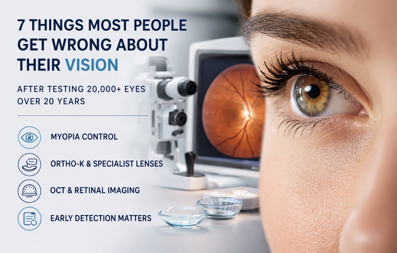
Optomap for Retinal Detachment Diagnosis
Optomap
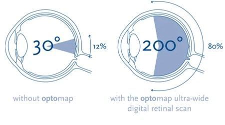
is an innovative technology that has streamlined the way retinal conditions are diagnosed, including retinal detachment: it's faster, more accurate, and more comfortable for patients compared to traditional methods. This advanced imaging technique provides a wide-field view of the retina, capturing more than 80% of its surface in a single image.
One of the most notable advantages of Optomap is that it doesn't require pupil dilation - as such, the process is quicker, more comfortable, and less invasive for patients. The detailed images produced by Optomap help eye doctors detect issues that might be missed with traditional methods.
Optomap employs low-power laser scanning to capture high-resolution images of the retina. The system uses two different lasers - one red and one green - to scan the retina. These lasers penetrate the different layers of the retina, capturing high-resolution images that can reveal issues like tears, detachments, and other abnormalities.
Key Features and Benefits
Optomap offers several key features and benefits that make it a superior choice for retinal imaging:
- Wide-Field Imaging: Optomap captures more than 80% of the retina in a single image, providing a comprehensive view that traditional methods can't match.
- Non-Invasive: The imaging process is quick, painless, and doesn't require pupil dilation, making it more comfortable for patients.
- Early Detection: The detailed images produced by Optomap help detect early signs of retinal conditions, allowing for timely treatment and better outcomes.
- Efficient Monitoring: Optomap images can be saved and compared over time, making it easier to monitor disease progression and treatment effectiveness.
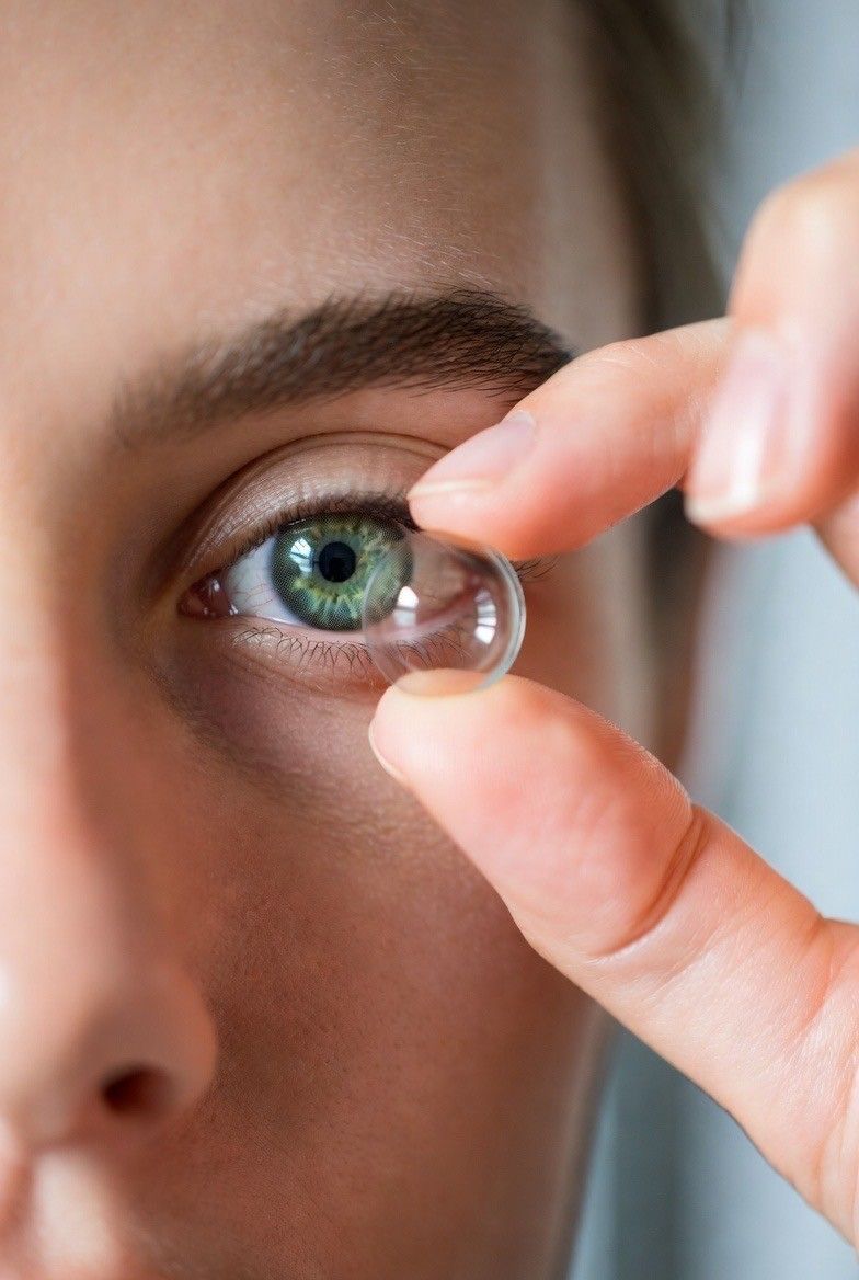
The Imaging Process
- Step 1: The patient looks into the Optomap instrument and focuses on an LED target.
- Step 2: The instrument uses low-power red and green lasers to scan the retina, capturing detailed images of its surface.
- Step 3: The images are displayed on a computer screen, where the eye doctor can examine them for signs of retinal detachment and other conditions.
The entire process takes just a few minutes and doesn't require any special preparation or recovery time.
The eye doctor will then look for specific signs that indicate retinal detachment, such as a separation between the retina and the underlying tissue. They will also examine the images for other issues that could affect vision, such as diabetic retinopathy, macular degeneration, and glaucoma.
Comparing Optomap with Traditional Methods
Compared to traditional methods like standard eye exams, pupil dilation, and ultrasound, Optomap offers several significant advantages:
- More Comprehensive View: Optomap captures a wider area of the retina, providing a more complete picture of its health.
- Greater Comfort: The non-invasive nature of Optomap imaging makes it more comfortable for patients, as it doesn't require pupil dilation.
- Faster Results: The imaging process is quick, allowing for faster diagnosis and treatment planning.
- Better Monitoring: Optomap images can be saved and compared over time, making it easier to track changes in retinal health.
Treatment Options for Retinal Detachment
Once retinal detachment is diagnosed, prompt treatment is essential to prevent permanent vision loss. Several treatment options are available, depending on the severity and location of the detachment.
Laser Surgery
Laser surgery, also known as photocoagulation, is a common treatment for retinal detachment. This procedure uses a laser to create small burns around the retinal tear, sealing it to the underlying tissue and preventing further detachment.
The procedure is usually performed in an outpatient setting and takes about 20 to 30 minutes. Patients may experience some discomfort and blurred vision for a few days after the surgery, but recovery is generally quick.
Pneumatic Retinopexy
Pneumatic retinopexy is another treatment option for retinal detachment that involves injecting a small gas bubble into the eye. The bubble presses against the detached retina, helping it reattach to the underlying tissue.
The patient must keep their head in a specific position for several days to ensure the bubble stays in place - the bubble eventually absorbs on its own, and the retina remains attached. This procedure is less invasive than other surgical options and has a shorter recovery time.
Choose Eyecare Opticians For Optomap Retinal Exam In Kingston Upon Thames

If you're looking for a reliable and comfortable way to monitor your retinal health, consider booking an Optomap retinal exam with Eyecare Opticians in Kingston Upon Thames. Our state-of-the-art technology and experienced eye care professionals ensure that you receive the best possible care.
Detecting retinal detachment and other eye conditions early is vital for safeguarding your vision - with Optomap, we provide you with a comprehensive and detailed assessment of your retina, identifying problems in their early stages and advising on the most suitable treatments.
You can resume your normal activities immediately after the exam, without the light sensitivity and blurred vision that typically follow pupil dilation. This makes Optomap an excellent choice for individuals who may find dilation particularly bothersome, such as children and those with busy schedules.
With Optomap, we save the images and compare them over time, which is particularly beneficial for patients with chronic eye conditions that require ongoing management. By providing a clear and consistent record of retinal health, Optomap helps our team make informed decisions about treatment and follow-up care.
Don't wait until you experience symptoms to take care of your eyes. Schedule an appointment with us today and take the first step towards maintaining a healthy vision for life!
Frequently Asked Questions (FAQ)
What is retinal detachment and how does it affect vision?
Retinal detachment occurs when the retina pulls away from its normal position at the back of the eye. This can cause symptoms such as flashes of light, floaters, and a shadow over part of the field of vision. If left untreated, it can lead to permanent vision loss.
How often should I get an Optomap retinal exam?
It's generally recommended to get an Optomap retinal exam annually, especially if you have risk factors for retinal detachment or other eye conditions. Your eye doctor may recommend more frequent exams based on your individual health needs.
Is Optomap imaging safe and painless?
Yes, Optomap imaging is both safe and painless: the process uses low-power lasers to capture detailed images of the retina without any discomfort or side effects. It's a quick and non-invasive procedure that doesn't require pupil dilation - and at Eyecare Opticians, we’re always ready to answer any of your questions to help you get the best vision possible!
Can Optomap detect other eye conditions as well?
Yes, Optomap can detect a range of eye conditions beyond retinal detachment, including diabetic retinopathy, age-related macular degeneration, glaucoma, and even cancer. The high-resolution images it provides allow for early detection and monitoring of these conditions.